|
|

The Visual Computing Forum, or VCF, is a
series of seminars organized by the visualization
group with selected talks from the fields of
visualization, image processing, computer graphics,
and so on. The individual seminars are arranged
approximately once a month, on Fridays from 11am to
12am, and they will be interleaved with the MedViz seminars.
They will be held either at the Høyteknologisenteret
or at the VilVite
Science Center. If you wish to be informed about upcomming VCF events, please write an e-mail to "vcf.bergen@gmail.com", "Helwig.Hauser@UIB.no" or "Sergej.Stoppel@UIB.no".

|
|
December 2011
Holidays finally!
The VCF goes on holyday as well: the next seminar will be on Friday
13th January and all the details will be announced soon on this page.
In the meantime the VCF staff wish you Merry Christmas and Happy New
Year!
|
|
|
09
December 2011
MedViz Seminar - Imaging and New Targets for
Personalised Medicine in Endometrial Cancer
Title:Imaging and New Targets for
Personalised Medicine in Endometrial Cancer
Speakers:
1. Helga B. Salvesen,
Department of Gynaecology, Haukeland University
Hospital
2. Ingfrid S. Haldorsen and Jenny
A. Husby, Department of Radiology, Haukeland
University Hospital
Check the
seminar page for the detailed abstract.
|
 |
|
02
December 2011
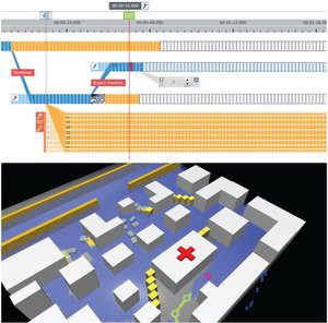
Visual
Steering to Support Decision Making in Visdom
Speaker: Eduard Gröller
Place: Konferanserom C (TM51:KONFC),
VilVite Science Center, Thormøhlensgate 51
Time: Friday 02 Dec 2011, from 11.00am to
12.00am
Abstract:
Computer simulation has
become an ubiquitous tool to investigate the
nature of systems. When steering a simulation,
users modify parameters to study their impact on
the simulation outcome. The ability to test
alternative options provides the basis for
interactive decision making. Increasingly complex
simulations are characterized by an intricate
interplay of many heterogeneous input and output
parameters. A steering concept that combines
simulation and visualization within a single,
comprehensive system is largely missing. This
talk targets the basic components of a novel
integrated steering system called Visdom to
support the user in the decision making process.
The proposed techniques enable users to examine
alternative scenarios without the need for
special simulation expertise. To accomplish this,
we propose World Lines as a management strategy
for multiple, related simulation runs. In a
dedicated view, users create and navigate through
many simulation runs. New decisions are included
through the concept of branching. To account for
uncertain knowledge about the input parameters,
we provide the ability to cover full parameter
distributions. Via multiple cursors, users
navigate a system of multiple linked views
through time and alternative scenarios. In this
way, the system supports comparative visual
analysis of many simulation runs. Since the
steering process generates a huge amount of
information, we employ the machine to support the
user in the search for explanations inside the
computed data. Visdom is built on top of a
data-flow network to provide a high level of
modularity. A decoupled meta-flow is in charge of
transmitting parameter changes from World Lines
to the affected dataflow nodes. To direct the
user attention to the most relevant parts, we
provide dynamic visualization inside the flow
diagram. The usefulness of the presented approach
is substantiated through case studies in the
field of flood management. The Visdom application
enables the design of a breach closure by
dropping sandbags in a virtual environment.
Additional material:
Flyer, Eduard
Gröller webpage,
The Visdom project
|
|
|
|
25
November 2011
MedViz
Seminar - Drug-delivery by Microbubbles
Title: Drug-delivery by Microbubbles
1. The physics and mechanics of sonoporation
2. Biological applications of sonoporation
3. How to obtain local drug delivery to the
pancreas
4. MedIm - Norwegian Research
School in Medical Imaging - a short presentasjon
Speakers:
1. Researcher Spiros
Kotopoulis (1)
2. Researcher Antony
Delalande (2)
3. Associate professor,
Georg Dimcevski, UiB (3 and 4)
Abstract:
Ultrasound is very well
known for its use in clinical diagnostics and
non-destructive testing. For past few years its
use for therapeutics has been explored. Existing
approved uses include physiotherapy, surgery using
high-intensity focus ultrasound and facial
rejuvenation.
Our work demonstrated the
manufacture of ultra-high resolution transducers
capable of thermal ablation with millimetre
accuracy. Such methods could be used to treat
areas where invasive surgery is not possible.
During high-intensity ultrasound, gas cavities may
form (acoustic cavitation), disrupting the
ultrasound propagation. This can affect the
efficiency of for high-intensity focused
ultrasound surgery. We designed and built a tool
to investigate and control cavitation to help
enhance the effect of ultrasound or therapy.
In clinical-diagnostic imaging, gas microspheres
(microbubbles) are used to increase the blood
acoustic backscatter. These microbubbles are also
acoustically active, allowing for complex acoustic
interactions. We took advantage of this and showed
that it is possible to precisely control and
manipulate thousands of microbubbles using
ultrasound in the clinical diagnostic range.
Recent research has also shown that microbubbles
in the presence of ultrasound have the ability to
enhance cellular drug or gene uptake. This is
known as sonoporation. We investigated this
phenomenon using HeLa cells and saw that there was
a specific threshold where this uptake was most
efficient. Using these ideal settings we showed
gene transfection in to mice tendons that lasted
for over 100 ays. This is also resulted in
restoration of the collagen fibers.
We
investigated the physical mechanism behind this
increased uptake and saw that it was possible to
direct a fluorescence-coated microbubble directly
into cells where it subsequently dissolved.
|
 |
|
04
November 2011
CMR
Computing - Visual computing and applications
Speaker: Ola Kristoffer Øye, Yngve
Heggelund (PhD, Senior scientists, CMR Computing)
Place: Konferanserom C (TM51:KONFC),
VilVite Science Center, Thormøhlensgate 51
Time: Friday 04 Nov 2011, from 11.00am to
12.00am
Abstract:
ANALYSIS AND
VISUALIZATION OF MARINE ACOUSTICS DATA FOR
ABUNDANCE ESTIMATION OF MARINE RESOURCES -
Marine acoustics is one of the primary data types
used by marine scientist for abundance estimation
of marine resources. Large amounts of
multi-frequency echosounder and sonar data is
regularly collected by research vessels, and
analyzing and extracting quantitative measures
from these data is crucial for good estimates of
fish stocks, which again leads to catch quotas
for the fisheries industry. The Institute of
Marine Research (IMR) and CMR have for several
years developed analysis and visualization
techniques for analyzing marine acoustics data.
The techniques and work flows has been
implemented in the application "LSSS - Large
Scale Survey System", which today is in use at
IMR and several other marine research
institutions around the world. The talk will
present some of the techniques, workflows and
features that has been developed.
INTERACTIVE
COMPUTATION AND VISUALIZATION OF WIND FARM FLOW
FIELDS BASED ON MODEL REDUCTION - Wind turbines
in wind farms generate wakes, which reduces the
power production of turbines downstream of other
turbines. CFD (Computation Fluid Dynamics) is the
best candidate for describing complex wake
effects, but the application of such models is
computationally very expensive which limits their
practical use. This presentation will outline a
method of reduced order modeling based on CFD.
This provides a much faster computation of the
flow field, which enables the user to make
changes of the positioning of turbines within a
wind farm and interactively observe the effects.
While a CFD simulation takes hours to complete,
the flow field can be computed within seconds in
the reduced order space.
Additional material:
Flyer, CMR
Computing webpage,
Slides CMRComputing,
Slides Wind Farms
|
|
|
|
14
October 2011
MedViz
Seminar - Bergen Stroke senter
Webpage: http://medviz.uib.no/
Topic: Bergen Stroke senter
Place: BBB, konferanserommet
Time: Friday 14 Oct 2011, from 12.00am to
1.00pm
|
 |
|
30
Semptember 2011
Assessing
brain connectivity using RS-fMRI and graph
theory in the context of open discovery science
Speaker: Professor Arvid Lundervold, MD,
PhD
Place: Store Auditorium, 2nd floor,
Høyteknologisenteret (data blokk),
Thormøhlensgate 55
Time: Friday 30 Sep 2011, from 11.00am to
12.00am
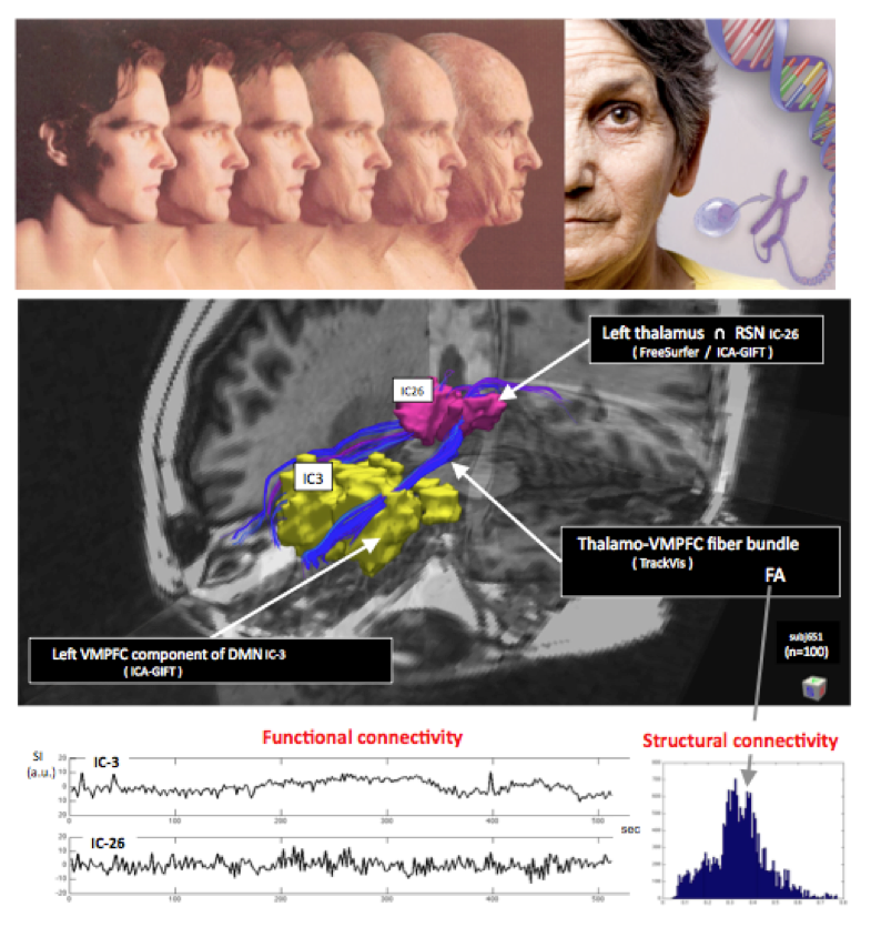
Abstract: The observation that spontaneous
BOLD fMRI activity is not random noise, but is
organized in the resting human brain as
functionally relevant resting state networks
(RSNs), has generated a new avenue in
neuroimaging and cognitive research - where brain
connectivity and graph theory are increasingly
important concepts for understanding and for
computation. One important application area of
this technology is the assessment of healthy
aging, mild cognitive impairment, and Alzheimer's
disease. In this talk I will present ongoing
research on brain connectivity and graph analysis
methodology applied to the aging brain of healthy
elderly people in the Bergen area as part of a
larger longitudinal study of cognitive aging,
where multimodal MRI examinations,
neuropsychological testing and genome wide
association (GWAS) data are included. In the
functional brain imaging part, two quite
different time scales are coming into play:
epochs of ~10 min resting state fMRI recordings
sampled at 0.5 Hz; and long-term changes in such
recordings over a period of ~3 years. The talk
will be put in the context of the recent paper by
B. Biswal et al. "Toward discovery science of
human brain function", PNAS
2010;107(10): 4734-4739, and the Norwegian
Academy of Science and Letters' Centre for
Advanced Study (CAS) 2011-2012 project "Cogniton
in aging - contributions of cognitive
neuroscience and cognitive neurogenetics", headed
by Prof. Ivar Reinvang, UiO
Additional material:
Flyer, Prof.
Lundervold webpage,
|
|
|
|
16
Semptember 2011
MedViz
Seminar
Official page: MedViz
seminar webpage
Speakers:
1. Introduction, Arvid
Lundervold, Professor UiB
2. MIC
organization / core facilities, Geir Olav
Løken, Senior Executive Officer, UiB
3. Services - imaging equipment, data storage
& retrieva, Hege Avsnes Dale, Chief Engineer,
UiB
4. Image analysis - report from an
application in progress, Erlend Hodneland,
Researcher, UiB
Place: Aud 4, BBB
Time: Friday 16 Sep 2011, from 12.00am to
1.00pm
Abstract: A core facility, such as MIC, is
a compilation of equipment and highly qualified
staff under a common organizational umbrella. Its
mission is dual. Firstly it carries out R&D on
the basis of its equipment in order to forward
its range and quality to users and ensure that
these are in the frontline of its field.
Secondly, available and newly developed methods
are implemented as service to be offered for the
benefit of the wider research environment at a
low price and without any demand for
collaboration. The Molecular Imaging Center
offers a wide range of services, ranging from
access to instrumentation and equipment via
courses and training to full service combining
sample preparation with image acquisition and
analysis carried out by our highly competent
staff. MIC is equipped for imaging at the
nanometer- to the micrometer and sub-millimeter
levels. This enables us to facilitate research
ranging from the molecule level, via cell
organelles and cells to whole animals. MIC is
thus a true translational core facility. In
addition to offering sample preparation / animal
handling at all levels we specifically have
equipment for electron-, fluorescence- and
confocal microscopy (including 2-photon), high
throughput imaging, magnetic resonance imaging,
optical imaging, and flow cytometry. MIC is
co-localized with an animal stable and has highly
qualified technical and scientific personnel
operating and maintaining all instruments. One of
the many ongoing project at MIC is related to
modeling melanoma brain metastasis, aiming at
more targeted treatment of cancer. In this
project the 7 T small animal scanner at MIC is
used to image metastatic lesions in the mouse
brain, where development of task specific
segmentation algorithms is necessary to make
large scale quantification across several animals
and experimental conditions feasible. This
automated image analyses approach, using the
MATLAB computing environment, will be presented
and discussed.
|
 |
|
2
Semptember 2011
Passing
Through the Trough of Disillusionment of
Illustrative Visualization
Speaker: Professor Ivan Viola
Place: Room 3137, Høyteknologisenteret
(data blokk), Thormøhlensgate 55
Time: Friday 2 Sep 2011, from 11.00am to
12.00am
Abstract: Efficient illustration craft is
a vast source of inspiration for development of
new visual abstractions in data visualization.
Many new illustration-inspired techniques have
emerged up to now, primarily arguing their
validity with a statement like: "The illustrators
have been using this technique for centuries and
therefore we adapt their technique for
interactive data display...". Argumentation of
such kind was stimulating in the initial phase of
illustrative visualization research, but nowadays
this reasoning is no longer satisfactory. It is
becoming apparent that ad-hoc adaptation can have
arbitrary outcome. A systematic adaptation
requires a vivid dialog with illustrators and a
well-founded reasoning by means of the vision and
cognitive sciences. This talk will assess the
efficiency of selected visual abstractions,
adapted for interactive visualization, in terms
of their consistency with established perceptual
principles.
Additional material:
Flyer,
Video, Slides
|
 VCF seminars in
2017,
2016,
2015,
2014,
2013,
2012,
2011
VCF seminars in
2017,
2016,
2015,
2014,
2013,
2012,
2011
|
|